admin 发布的文章

Fluoroscopic Angiography Assessment, Protocols, and Interpretation

Excerpt 摘录
Fluoroscopy-guided catheter angiography is an interventional procedure that uses percutaneous access of arteries with needles and catheters to inject contrast for vessel opacification. This procedure may be diagnostic or therapeutic. While some providers use angiography as a general term to include visualization of arteries, veins, or lymphatics, this article uses the term angiography to refer solely to the visualization of arteries, also known as arteriography. The terms venography and lymphangiography refer to veins and lymphatic vessels, respectively. Nevertheless, the principles behind angiography are widely applicable to other vessel types.

Sven Ivar Seldinger’s discovery of a technique in 1953 which described the substitution of a needle or trocar by a percutaneous catheter, since called the Seldinger technique, allowed for the possibility of catheter angiography and the birth of Interventional Radiology as a specialty. The following decades gave rise to several non-invasive angiographic advancements, including computed tomographic and magnetic resonance angiography.

Catheter angiography remains the gold-standard for a wide variety of pathologies. Today, catheter angiography is used to interrogate arteries in nearly every part of the human body, including the brain, neck, heart, chest, abdomen, pelvis, and extremities. Applications of catheter angiography are immense and include identification of arteriovenous malformations, aneurysms, atherosclerosis, embolisms, dissections, congenital abnormalities, stenosis, hemorrhage, and other arterial pathologies. Angiography may help guide implantation of stents, grafts, or provide assessment before surgery, chemoembolization, or internal radiation therapy. Fluoroscopy is undoubtedly the most important tool for an interventionalist. Herein, the general principles of fluoroscopy-guided catheter angiography are described, with a focus on the appropriate use of fluoroscopy-guidance for the diagnosis of arterial pathology.

[FROM]
Brian Covello 1, Brett McKeon
In: StatPearls [Internet]. Treasure Island (FL): StatPearls Publishing; 2024 Jan.
2023 Feb 13.
Affiliations Expand
PMID: 33760526 Bookshelf ID: NBK568767
[/FROM]

angiography 血管造影
arteriography 动脉造影
venography 静脉造影
lymphangiography 淋巴管造影

https://tumourclassification.iarc.who.int/welcome/

WHO Classification of Tumours Online presents the authoritative content of the renowned classification series in a convenient digital format. Now combining the fourteen most recent volumes of the series in a searchable format, with high quality images and whole slide images.
WHO Classification of Tumours Online is indispensable for pathologists and cancer specialists worldwide. New volumes will be added regularly, ensuring immediate access to the latest content.

脑膜瘤病理类型多样,临床表现复杂,是中枢神经系统最常见的原发肿瘤。大多数良性脑膜瘤(WHO 1级)患者通过手术和/或放射治疗能够获得良好临床预后。然而,部分具有进展趋势的良性脑膜瘤和不典型或恶性脑膜瘤(WHO 2-3级)仍面临着复发率高,后期治疗困难等问题,严重影响患者生存质量。近年来,越来越多研究发现脑膜瘤从基因、蛋白到代谢等各个层面的分子特征与其病理分级、患者临床预后密切相关;此外,仅依据组织形态作为主要判定标准的传统病理分级和分型已不足以准确预测所有脑膜瘤患者的临床预后。2021年,第五版中枢神经系统肿瘤分类分级指南(WHO CNS5)更新了部分中枢神经系统肿瘤亚型的命名和分级方法,并首次将脑膜瘤分子特征纳入诊断标准,但仍缺乏有关问题的系统阐述。

因此,在2021年5月初,在国家神经疾病医学中心、复旦大学附属华山医院周良辅院士、毛颖院长的指导下,由华山医院的宫晔教授和钟平教授发起、启动了这部共识的筹备工作,并在国际临床实践指南注册平台完成了注册(IPGRP-2022CN234)。同年11月,共识编写委员会正式成立,共汇集了来自全国各地主要神经外科单位的25名神经外科专家,包括中华医学会神经外科学分会主任委员、候任主任委员、副主任委员及6位常委,其余专家也都是全国委员和全国青委委员;并邀请华山医院神经病理室的陈宏教授;复旦大学循证医学中心的陈世耀教授团队,兰州大学循证医学中心的陈耀龙教授团队共同参与共识修订。同时设立秘书组筹备和协调编写工作;并携手CMJ编辑部召开共识编写项目线上启动会、修订会和定稿会。历经制定标准、文献调研、临床问题构建、确定共识框架、撰写和讨论修改文稿、评分投票、修稿审定等环节,最终全部编写完成。

英文版全文链接
https://journals.lww.com/cmj/Fulltext/2022/08200/Molecular_diagnosis_and_treatment_of_meningiomas_.2.aspx

中文版全文链接
https://rs.yiigle.com/CN112050202304/1458625.htm

通常把蝶窦向翼突根方向气化形成的位于翼管-圆孔连线(V-R line)之外的空间,称作蝶窦外侧隐窝(lateral recess of sphenoid sinus,LRSS)。

LRSS位于颅底,位置较深,与周边血管、神经关系复杂,是自发性脑脊液漏和脑膜脑膨出的高发区域,手术操作具有挑战性。

常用的传统手术入路包括:经颅入路、经面部入路和经口咽入路。

随着鼻内镜技术的不断拓展,使用经鼻经内镜入路技术处理上述颅底病变已日趋成熟,根据颅底疾病的具体侵袭范围可综合选择经鼻经斜坡入路、经鼻经蝶窦入路、经鼻经眶入路和经鼻经翼突入路等,其中经鼻经翼突入路术式的应用和报道目前较少见。

解剖结构
蝶窦只存在于灵长类动物,蝶窦向周围空间各个方向的气化程度变异很大,使得蝶窦与视神经、海绵窦、颈内动脉、垂体、第Ⅲ~Ⅵ对脑神经以及腹侧脑干的解剖关系异常复杂。蝶窦向翼突根及圆孔侧方气化形成蝶窦外侧隐窝,根据其在冠位层面由内向外的气化程度将其分为3型:气化不超过翼管为Ⅰ型(25%),即无侧隐窝型;气化超过翼管但不超过圆孔为Ⅱ型(39%);气化超过圆孔为Ⅲ型(37%),常导致中颅窝骨板变薄,见图4。
640.jpg
图4 蝶窦外侧隐窝的分型
4a:气化不超过翼管为Ⅰ型;
4b:气化超过翼管但不超过圆孔为Ⅱ型;
4c:气化超过圆孔为Ⅲ型。

翼突是蝶骨的一部分,分为翼突根部、内板和外板;以翼突根部为中心,其前方为翼腭窝,上方为眶上裂,内上侧为蝶骨体,外上为蝶骨大翼根部,外下为颞下窝,后方与岩尖关系密切;翼突根部内侧的骨孔为翼管前部开口,其内的翼管神经、翼管动脉出管口后向外下走形进入翼腭窝,翼管的后部开口恰位于颈动脉沟的下缘下,翼管以及其内翼管神经和动脉是颈内动脉岩骨段前膝部或破裂孔段的重要标志。翼管前部开口的外上为圆孔,其内走行的V2起源于中颅窝Meckle腔的三叉神经节,穿圆孔向前进入翼腭窝,随后向内下发侧支进入蝶腭神经节,其主干向外上翻折入眶下裂进入眶下神经管;翼外板根部的稍外侧为卵圆孔,V3向外下穿卵圆孔入颞下窝;翼突内、外板之间为翼窝,翼窝内包含翼内肌起始部,其后上部的卵圆形凹陷为舟状窝,提示咽鼓管的翼突部。

翼突已成为颅底手术通道的枢纽区域,对于处理前方的翼腭窝,外侧的颞下窝后部的蝶窦外侧隐窝、Meckle腔、外侧海绵窦,外侧的中颅窝、岩尖、岩斜区,上方的眶上裂、眶尖,以及下方的咽鼓管区病变都有重要意义。

手术入路
颅底区域的手术主要有经颅入路(包括经颞骨-颅中窝入路、经迷路入路、经颞下窝入路、经后颅窝入路等)、经面部入路、经口咽入路和内镜经鼻入路等路径。相较之下,内镜经鼻路径具有更小进路、更大范围切除和更广阔手术视野等特点。

根据不同疾病在颅底的侵犯范围不同,Schwartz等将内镜经鼻入路术式详细划分为4种主入路及若干亚路径,包括:

①经鼻入路:经筛板、经斜坡、经齿突路径;

②经蝶窦入路:经蝶鞍、经鞍结节、经蝶骨平台、经斜坡、经海绵窦路径;

③经筛窦入路:经筛凹、经眶、经蝶窦路径;

④经上颌窦入路:经翼突路径。

对于蝶窦外侧隐窝的病变(例如脑脊液鼻漏、脑膜脑膨出等),尤其第Ⅲ型且位于蝶窦外侧隐窝前内侧壁的病变,由于内镜技术及鼻腔通路的角度限制,单纯经蝶窦入路较难将该区域病灶完全清除。经翼突入路通过磨除翼突根前壁,以直视蝶窦外侧隐窝,可完整处理整个蝶窦外侧隐窝的病变,常联合经蝶窦入路共同处理范围较大的颅底疾病。

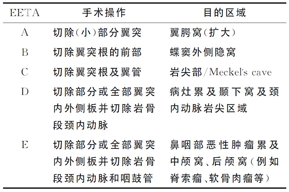
Kasemsiri等把经翼突入路能到达的鼻颅底区域划分成5个部分,并进一步将翼突入路术式根据暴露范围分为5种亚型(表2)。
650.jpg

翼管和圆孔是经翼突路径术式最重要的解剖标志。Karci等通过影像学测量发现圆孔始终位于翼管的上外侧,翼管与圆孔的距离为8.50~10.50mm,平均(9.45±0.60)mm,翼管到蝶腭孔距离为5.5~7.0mm,平均(6.30±0.47)mm。翼管神经和动脉是指示颈内动脉岩骨段前膝部(破裂孔段)的重要标志,V2已成为评估眶上裂和海绵窦的下界以及Meckle腔和颅中窝的上界的手术标志。

随着内镜、影像学技术的不断发展,对解剖认识的逐渐深入,新衍生术式也在不断被提出。Patel等提出经对侧上颌骨入路处理岩尖区包括胆脂瘤、软骨肉瘤等疾病,可减少对颈内动脉的操作和保留翼管神经。Truong等提出采取上颌窦根治术入路经上颌窦经蝶骨大翼路径处理中颅窝包括Meckle腔、海绵窦、翼腭窝、颞下窝等部位的疾病。

手术方式:患者取卧位,用1%注射用盐酸丁卡因20ml和0.1%肾上腺素2ml浸湿棉片行表面麻醉,5min一次,共3次。术中见蝶筛隐窝处可见搏动性清水样溢液,清除后可见蝶窦口黏膜肿胀,结合术前影像学检查,判断为蝶窦外侧隐窝的脑脊液鼻漏可能。切除患侧钩突充分开放上颌窦,开放前后组筛窦,向上暴露筛顶,向外暴露眶纸样板。去除蝶窦前壁,充分开放蝶窦,显露蝶窦内重要解剖标志,如鞍底、斜坡隐窝、颈内动脉隆突、视神经隆突等,术中证实病变位于蝶窦外侧隐窝,有搏动性的脑脊液鼻漏,手术操作困难。遂自上颌窦开口向后将上颌窦内侧壁部分骨质去除至蝶腭孔前方,以Kerrison咬骨钳咬除部分上颌窦后壁,并咬除筛嵴,磨除腭骨垂直板前上部,充分暴露翼腭窝内容物,包括脂肪、神经、血管及结缔组织,电凝烧灼蝶腭动脉。使上颌窦、后筛以及蝶窦相融合于翼腭窝表面,结合术前阅片定位翼管神经位置,多数情况下需要行翼管神经和动脉电凝后切断,便于进一步暴露,将翼腭窝内容物向外下分离,电凝上颌动脉及其分支,充分暴露翼突根部的骨质并磨除,以较好地显露蝶窦侧隐窝结构,结合术前阅片结果注意识别外侧壁上在翼管神经血管根部外上方呈水平走行的三叉神经上颌支(V2)隆起。接着清理蝶窦侧隐窝病变(1例为膨出的脑膜脑组织,1例为真菌侵袭性病灶),结合术前影像学资料在内镜下定位颅底骨质缺损,行脑脊液鼻漏的修补。取患者部分大腿股四头肌肌肉组织(捣成肌浆)及适当大小阔筋膜以“三明治法”修补2例,鼻中隔黏骨膜瓣单层修补1例及阔筋膜内置及鼻中隔黏骨膜瓣外贴双层修补1例,明胶海绵颗粒填塞,碘仿纱条固定,术后2周拔除碘仿纱条。

Hyperostotic Invasive Meningioma of the Sphenoid Ridge Surgical Considerations: 2-Dimensional Operative Video
Essayed, Walid Ibn MD; Aboud, Emad MD‡; Al-Mefty, Ossama MD
Author Information
*Department of Neurosurgery, Brigham and Women's Hospital, Harvard School of Medicine, Boston, Massachusetts, USA;

‡Department of Neurosurgery, Arkansas Neuroscience Institute, Little Rock, Arkansas, USA

Correspondence: Ossama Al-Mefty, MD, Department of Neurosurgery, Brigham and Women's Hospital, Harvard Medical School, 75 Francis St, Boston, MA02115, USA. Email: almeftyossama@bwh.harvard.edu

Operative Neurosurgery 22(3):p e124-e125, March 2022. | DOI: 10.1227/ONS.0000000000000074

Hyperostotic invasive sphenoid ridge meningiomas are a distinct and recognized entity.1-4

They are characterized by extensive bone involvement and expansive “en plaque” intradural extension.3,5 The goal of this surgery is improving and preserving the visual function and ocular motility while esthetically correcting and reconstructing the exophthalmos and temporal deformity.4,5 The surgery includes radical removal of the involved bone of the sphenoid ridge, orbital walls, and middle fossa floor, in addition to the “en plaque” dural and periorbital involvement and intradural extension.3,5,6 Early decompression of the optic nerve and orbital elements allows preventing visual function deterioration and yields frequent improvement. Thorough planned reconstruction can be achieved by molding a malleable mesh to the patient's skull before the extensive bone removal. Careful orbital wall reconstruction allows one to achieve a good postoperative cosmesis and prevent enophthalmos.4 We demonstrate the principles of this surgery through 2 cases of 46-year-old and 53-year-old women. The first case demonstrates the importance of middle fossa extensive drilling of involved bone and reconstruction while the second case demonstrates the visual decompression and postoperative improvement. The patients consented to the surgeries and publication of their images.
Sphenoid Ridge Meningioma